
Sacroiliac Joint
Sacroiliac Joint Fusion in Oklahoma City
Minimally invasive sacroiliac joint fusion stabilizes the SI joint in patients whose low back and buttock pain has been traced to dysfunction, degeneration, or instability of the sacroiliac joint and who have not responded adequately to conservative treatment.
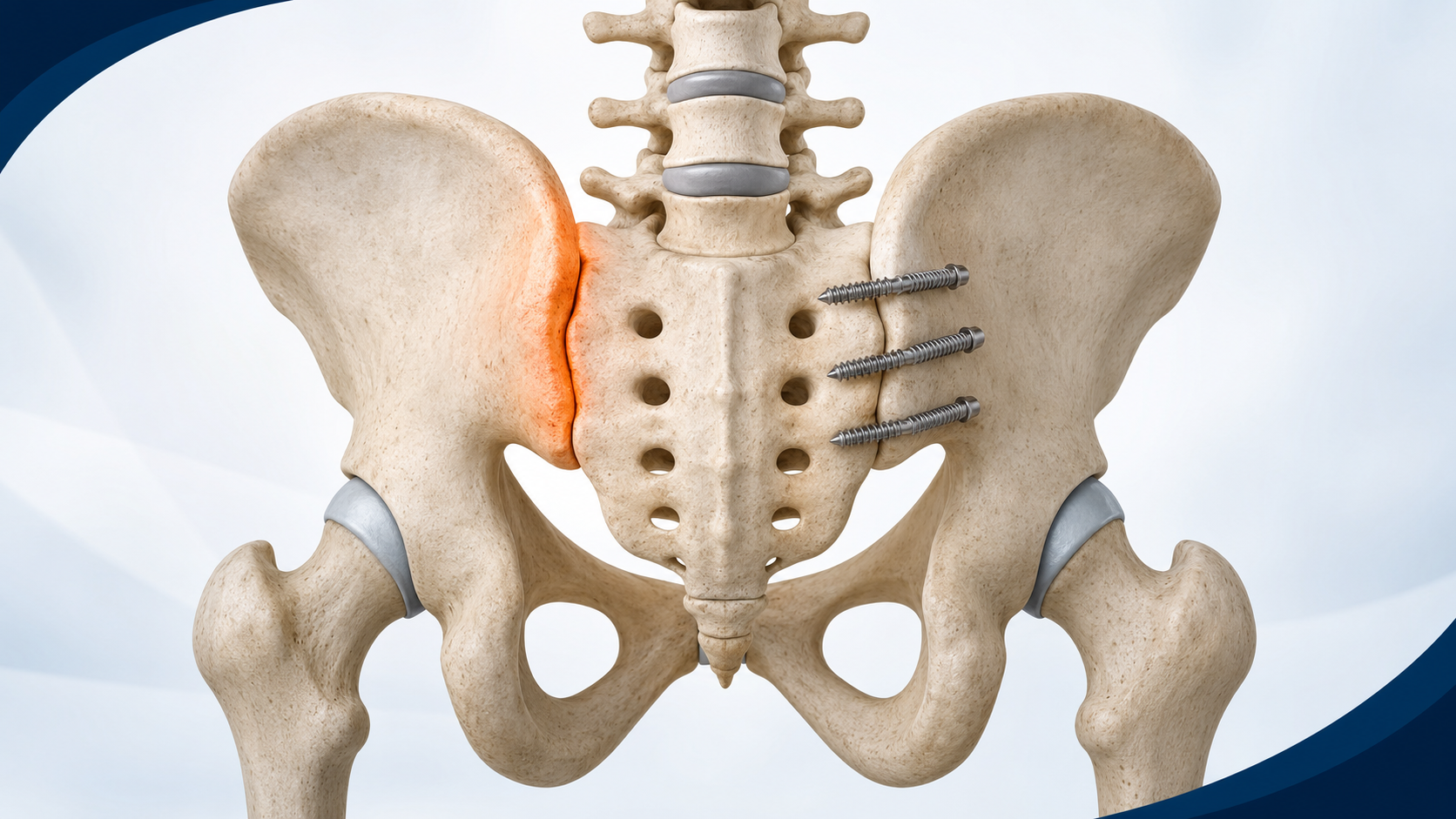
What this image shows
- The highlighted side demonstrates the location of a symptomatic sacroiliac joint.
- The opposite side shows generic minimally invasive implants stabilizing the joint across the ilium and sacrum.
- Fusion is considered only after the SI joint has been confirmed as the pain source and nonsurgical options have been addressed.
SI Joint Pain vs. Lumbar Spine Pathology
Patients searching for answers to "pain below the beltline radiating into the buttock" or "SI joint pain vs lumbar disc herniation" are often experiencing one of the most diagnostically challenging presentations in spine and orthopedic care. The sacroiliac joint, lumbar discs, and hip can all produce overlapping pain patterns — and the correct treatment depends entirely on identifying the correct source.
SI joint dysfunction is frequently misdiagnosed as lumbar pathology because both conditions cause low back and buttock pain. The distinguishing features are subtle: SI joint pain tends to be below the beltline, one-sided, and directly over the posterior SI joint, while lumbar disc pain more commonly produces dermatomal radiation into the leg below the knee. A fluoroscopic diagnostic injection into the SI joint remains the most reliable confirmation tool before any surgical decision is made.
SI joint dysfunction
Pain below the beltline, over the posterior dimple, worsened by sitting-to-standing transitions and one-legged stance.
Lumbar disc herniation
Dermatomal radiation below the knee, often with numbness or weakness in a specific nerve root distribution.
Hip pathology
Groin-predominant pain, limited internal rotation, worsened by weight-bearing — often confused with SI joint or lumbar sources.
Symptoms of SI Joint Dysfunction
- Low back pain localized to one side, just below the beltline
- Pain that radiates into the buttock, hip, or upper thigh
- Pain with prolonged sitting, standing, or walking
- Difficulty transitioning from sitting to standing
- Pain that worsens when bearing weight on one leg
- Tenderness directly over the SI joint
Confirming the Diagnosis
History and physical examination
Provocative tests that stress the SI joint help distinguish SI joint pain from lumbar spine or hip pathology.
Imaging
X-rays and CT scan assess joint degeneration, instability, and anatomy. MRI may be used to evaluate surrounding structures.
Diagnostic injection
A fluoroscopically guided injection of local anesthetic into the SI joint is the most reliable way to confirm the joint as the pain source. Significant relief following injection supports the diagnosis.
The Procedure
Minimally invasive SI joint fusion is performed through a small lateral incision. Implants are placed across the SI joint under fluoroscopic guidance to stabilize the joint and promote fusion. The procedure typically takes less than an hour and is performed on an outpatient or short-stay basis.
Patient selection is critical. Surgery is appropriate for patients who have confirmed SI joint pain on diagnostic injection, have not responded to conservative treatment including physical therapy and PRP injections, and have no other structural cause for their symptoms — such as lumbar disc herniation or lumbar spinal stenosis — that would better explain the clinical picture.
Schedule a Consultation
Call the office or submit a request online. We will follow up within one business day.